Stem cell therapy for the spine, before you consent to a fusion.

The epidural wore off. The surgeon has named the level and the operation, and you are not ready to sign. The physician team at Hospital Angeles in Tijuana, Mexico, 25 to 30 minutes from San Diego, reads your MRI and tells you plainly whether cell therapy fits your back or neck, or whether the surgeon is right. Spine cases get the most careful evaluation of any region we treat.

A patient coordinator typically calls within 1 business day. About 15 minutes, no cost, no obligation.

Medically reviewed by the physician team at Hospital Angeles · October 2026

Fluoroscopy, every level

A disc or facet sits deep, behind bone. The needle is placed under live X-ray and contrast dye confirms the level before any cells are released.

1 day, 1 session

Over the border and back, most patients home the same day. Follow-up calls at 1 week, then at 1, 3 and 6 months, then once a year; whether a second treatment makes sense is decided at those reviews, not sold up front.

Who qualifies, and who we send to a surgeon.

If you are reading this with your MRI report open in another tab, this is the section to read it against. Cortisone bought you a few months. An epidural bought a few more, then wore off. Then an ablation, and now a surgeon naming a level. That is not your back failing: a steroid calms inflammation for a while and puts nothing into the disc. The patients who do well here have an identifiable disc, facet, or nerve-root finding on MRI, a pain pattern that matches it, and a history like that one. The evaluation exists to separate those cases from the rest before anyone commits.

A physician in navy scrubs, seen from behind, reading a lumbar spine MRI on a lightbox and resting a finger on one disc
  • Not a fit

    Instability, or a nerve getting weaker.

    When a segment is unstable or a nerve is losing strength, decompression or fusion is the right tool, and we say so. Sudden weakness, numbness, or loss of bladder or bowel control is an emergency, not an evaluation.

  • Not a fit

    A large extruded herniation pressing on a nerve.

    A fragment of disc that has broken free and is causing a worsening nerve problem is a surgical case, routed to a surgeon promptly. A contained herniation is a different picture.

  • Strong fit

    Early disc disease with the disc height still there.

    MRI-confirmed disc pathology at 1 or 2 levels with preserved disc height, Modic changes (inflammation in the bone beside the disc) or a positive discogram, and a pain pattern that matches the findings. The profile we accept most often.

  • Also a fit

    A back that never settled after surgery.

    Persistent pain after a decompression, adjacent-level degeneration above or below a fusion, pain that came back after an ablation. The hardware is not the problem; what matters is whether the level driving your pain is identifiable and reachable.

  • Also a fit

    Nerve-root pain from a contained disc.

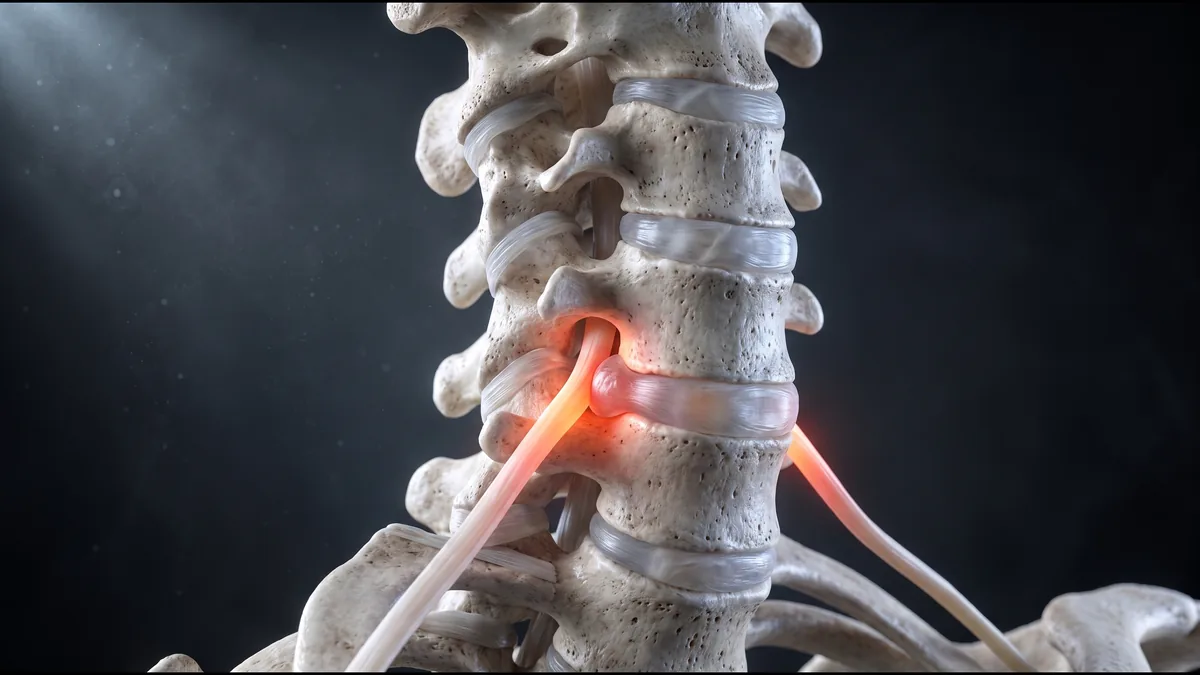
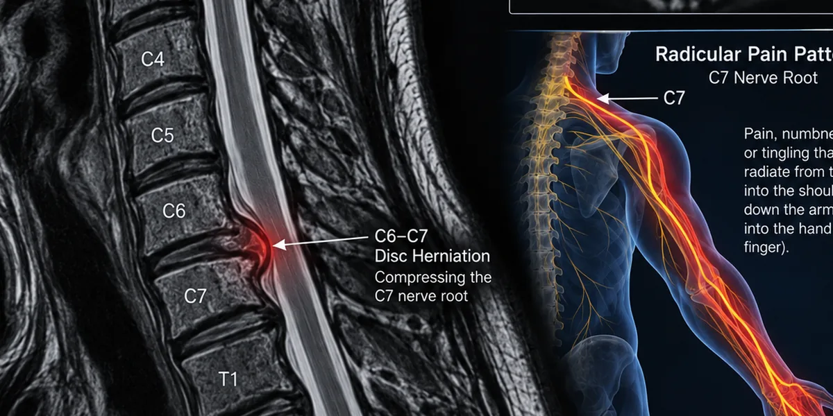
    Lumbar or cervical radicular pain (pain that runs down a leg or an arm from an irritated nerve root) from a contained disc or an inflamed root, at a level that matches the MRI.

  • Also a fit

    Facet pain confirmed by a block.

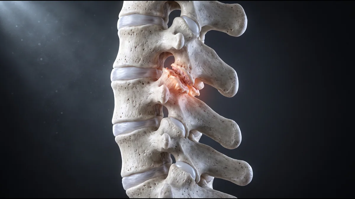
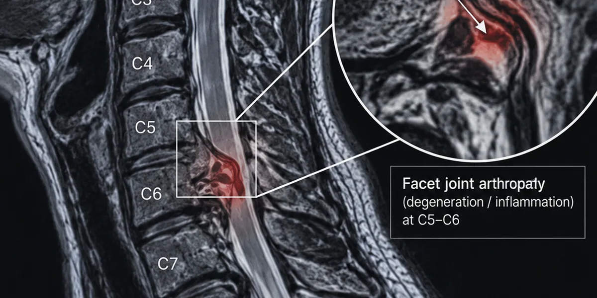
    Facet arthropathy (wear in the small joints at the back of each vertebra), lumbar or cervical, confirmed by a diagnostic block.

Your imaging decides. Not a sales team. Your first call is with a patient coordinator, who collects your MRI report for the physician team to read. Nobody quotes or books treatment before that read.

Book a call →

Spine injections under live fluoroscopy, at every level.

A disc, a facet joint, and a nerve root sit deep, behind bone. Ultrasound cannot see through bone. Fluoroscopy (a live X-ray) shows the vertebrae, the needle, and the contrast dye that proves the needle is at the right level before a single cell is released. No ultrasound shortcuts. No blind injection.

  1. Levels set from the MRI

    Read first, booked second.

    Disc height, Modic changes, facet degeneration, and what is pressing on which nerve root are read from your imaging against your pain pattern and any prior diagnostic blocks. Candidacy and levels come from that read.

  2. Local anesthetic, sedation available

    Still, comfortable, no general anesthesia.

    Local anesthetic always, with twilight sedation available if you want it, so the physician can place the full dose at each level. About 15 to 30 minutes on the table, depending on how many levels are treated.

  3. Needle under live X-ray

    The dye confirms the level. Then the cells.

    The physician confirms the needle is in the disc, the facet, or beside the nerve root at the chosen level, and only then releases the dose. Then the next level. 1 to 3 levels in a session.

  4. The cells, and what rides alongside

    Matched to your case, not pulled off a shelf.

    Bone-marrow and umbilical-cord MSCs (mesenchymal stem cells), often together, sometimes with chondrocytes (cartilage cells) when the disc or facet surface is part of the picture. PRP (platelet-rich plasma, from your own blood) or medical ozone are added in some cases, never in every one. Almost every joint and spine patient also receives a systemic IV the same day. Which cells, what dose, which levels: the physician team decides from your imaging.

Local soreness for about a week afterward is normal. The needle work creates inflammation by design, and that settles before anything else begins. Any change is measured over months, not days.

Degenerative disc disease and a herniated disc are two different cases.

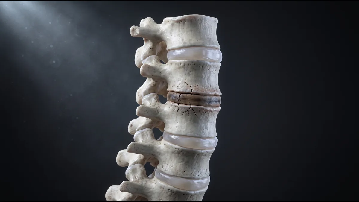
Degenerative disc disease is a disc drying and thinning over years. A herniation is disc material pushing out of place, and the kind of herniation decides everything.

Scott M., 68 · Degenerative disc

Degenerative disc disease

What the MRI has to show

  • Disc pathology at 1 or 2 levels, with the disc height preserved. A disc that has lost its height is read as a different case.
  • Modic changes (inflammation in the bone beside the disc), or a positive discogram.
  • A pain pattern that matches the level, and injections or an ablation that never held.
  • Cells go into the disc, or around it, at the level named, under fluoroscopy with contrast.
Illustration of two vertebrae with the disc between them dried, cracked and darkened, the bone on either side inflamed, but the disc still holding its height

Herniated disc

Contained, or extruded

  • A large extruded herniation causing a worsening nerve problem is a surgical case, and we route it to a surgeon promptly.
  • A contained herniation, including one driving chronic radicular pain, is evaluated individually from your MRI.
  • The target is the inflammatory environment around the nerve root and the disc, not the fragment itself.
  • Nerve regeneration is not the mechanism, and we do not promise it.
Illustration of two vertebrae with the disc between them bulging out at the back through a tear in its outer ring, the bulge still attached to the disc

Send the report and the images. The physician team reads them before anyone quotes or books you.

Book a call →

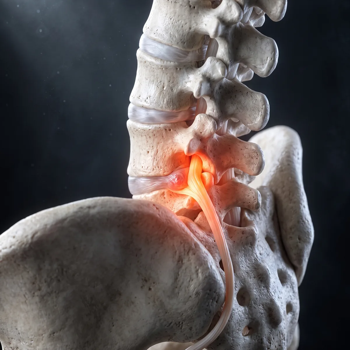
Sciatica is a symptom. We evaluate what is driving it.

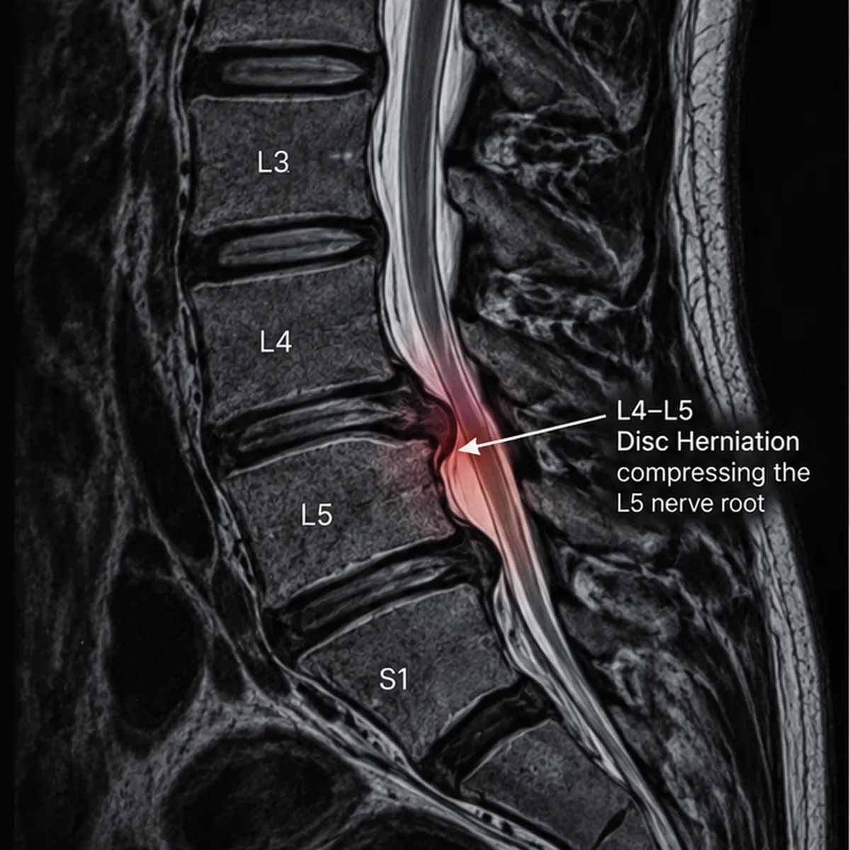
Pain that runs from the low back down the leg is a nerve root being irritated, by a disc or by the inflamed tissue around the root. The evaluation finds the level and the driver. The injection goes to the inflammatory environment around that root, under fluoroscopy, with contrast confirming the level. Nerve regrowth is not what we target and not something we promise. If something needs urgent care instead, we recognize it and route you there.

If your sciatica has a level on the MRI, L4, L5, or S1, send the report. The physician team reads it for the level before anyone books you.

Illustration of the lower spine and pelvis with one nerve root, where it leaves the spine, lit red where the disc presses on it, running down toward the legIllustrated lumbar MRI, sagittal view, with the disc at L4 to L5 pressing toward the nerve root

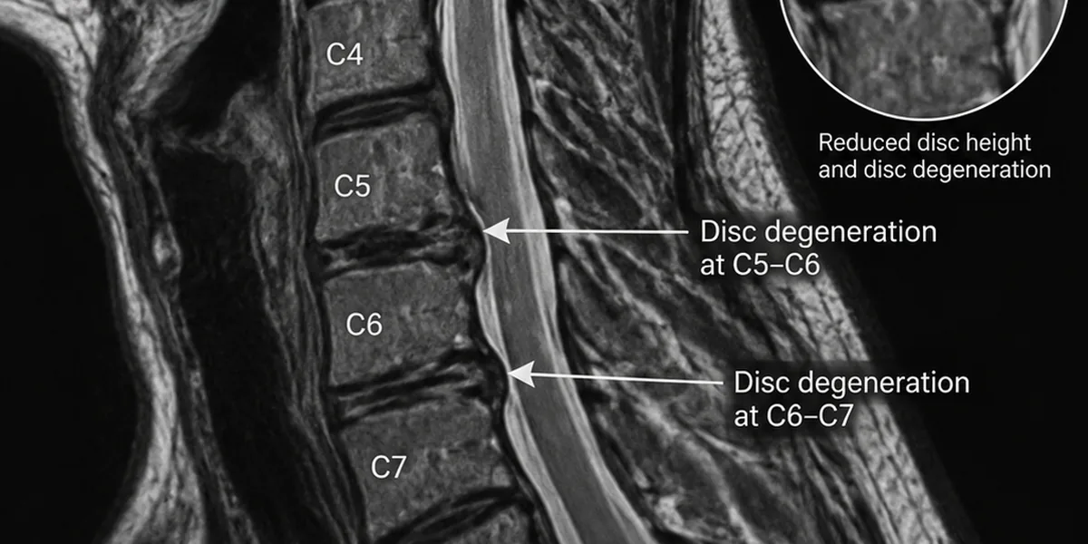
Neck pain and cervical discs, read the same way.

The cervical spine is evaluated exactly as the lumbar spine is: MRI read against your pain pattern, the level selected deliberately, fluoroscopic delivery with contrast confirmation.

Illustration of the cervical spine with one disc dried and darkened between two vertebrae, still holding its heightIllustrated cervical MRI, sagittal view, with the disc at C5 to C6 darkened and thinned

Disc degeneration in the neck.

A worn cervical disc at 1 or 2 levels, on the same criteria as the low back: disc height preserved, findings that match the pain.

Illustration of the cervical spine from behind, one small facet joint between two vertebrae worn and lit redIllustrated cervical MRI, sagittal view, with the facet joint at C5 to C6 marked

Facet pain in the neck.

Cervical facet arthropathy confirmed by a diagnostic block.

Illustration of the cervical spine with one nerve root, where it leaves the spine, lit red and running down toward the armIllustrated cervical MRI, sagittal view, with a disc at C6 to C7 pressing toward the nerve root

Radicular pain into the arm.

Nerve-root pain running into the shoulder, arm, or hand from a contained cervical disc or an inflamed root, at a level that matches the imaging.

Stem cells at the level, or a fusion across it.

"The back specialist told me that he wanted to fuse me." "I just didn't want to have bolts in my back. I just didn't want hardware." Scott M., a Celva patient, on the fork most people reading this are standing at. Here is the comparison, plainly.

Sally W., 64 · Back pain after a fusion

A fusion

Settles one segment, permanently

  • Hardware locks the level. Part of a structure built to move as a whole goes stiff, and the levels above and below take the extra load.
  • We often see those patients a year or two later with new pain somewhere else.
  • The right tool when a segment is unstable, a nerve is getting weaker, or a fragment of disc is pressing on it. Then we recommend a surgeon we trust.

Stem cells, at the level

Nothing removed, nothing fused

  • A fluoroscopy-guided injection into the disc, around it, into the facet, or beside the nerve root, at 1 to 3 levels.
  • Designed to calm the inflammatory environment around the disc and the nerve root through paracrine signaling (the cells' chemical messaging to nearby tissue).
  • 15 to 30 minutes on the table, under local anesthetic with twilight sedation available. Most patients home the same day.
  • One session. Whether a second treatment makes sense is decided at your follow-up reviews, not sold up front.
  • Not the right tool for instability or a worsening nerve deficit, and we say so.

The honest limit: the spine is the slowest region we treat to respond. The first days are soreness, and nothing is judged before the 1-month call.

Book a call →

Most patients sleep in their own bed that night.

Rewind to the pickup. Here is the day, start to finish, and where it can run long.

  1. Pickup, Southern California

    You, and a companion if you want one.

    A Celva driver collects you anywhere in Southern California and takes you home afterward. A companion is welcome, never required. 25 to 30 minutes from San Diego to Hospital Angeles, Tijuana, a 116-bed private hospital in the Angeles Health network.

  2. The physician, in person

    Before anything is injected.

    You meet the physician first. Your MRI has already been read; if the team wants a fresh look at the level, the hospital's own MRI can image it that day, with the radiologist's read the same day.

  3. On the table

    15 to 30 minutes.

    Local anesthetic, twilight sedation available, fluoroscopy in the room, contrast at every level, 1 to 3 levels. Almost every joint and spine patient also receives a systemic IV the same day.

  4. Discharge

    Most patients home the same day.

    The attending physician authorizes discharge, and most patients go home the same day. If your case calls for a night in Tijuana, you hear it during intake, not on arrival. Expect soreness in the spine where the needle went in, for about a week, then normal activity. Physical therapy is recommended, starting after the first 7 to 10 days.

After that: a pain-and-safety call at 1 week, then calls at 1, 3 and 6 months and once a year after that, and the spine is judged on the later ones because it takes longer to show anything. The physicians on every chart: Dr. Cesar Amescua (Medical Director), Dr. Rocio Ambrosio Nuño (anesthesiology and sedation), and Dr. Alejandro Castillo (interventional radiology).

Book a call →

"My kids would want to play with me, and I couldn't."

"I'd have to lay down because I'd be in pain." Jon C. hurt his back years ago, then re-sprained it doing jiu-jitsu: "weird, odd pain, numbness, tingling." Two flare-ups where he could not walk well. Doctors, MRIs, physical therapy. Surgery was the other option on the table. This is his film, in his own words, shot at Hospital Angeles on his treatment day. No script. Individual results vary.

Jon C. · Back pain

Spine-specific questions.

The ones we hear on almost every first call about a back or a neck.

Book a call →
Can stem cell therapy help me avoid a spinal fusion?
For discogenic, facet-mediated, or radicular (nerve-root) pain without instability, it is a legitimate path to evaluate before you consent to a fusion. When the picture points to surgery instead, like instability or a worsening nerve problem, fusion or decompression is typically the right answer, and we will tell you so.
Will this help sciatica or nerve pain down the leg?
Sciatica is a symptom; the structural and inflammatory driver is what we evaluate. The mechanism we target is the inflammatory environment around the nerve root, not nerve regeneration, and we do not promise nerve regrowth. Individual results vary.
Why fluoroscopy and not ultrasound for the spine?
A disc, a facet joint, and a nerve root sit deep, behind bone. Ultrasound cannot see through bone; fluoroscopy (a live X-ray) shows the vertebrae, the needle, and the contrast dye that proves the needle is at the right level before a single cell is released. For a shoulder tendon near the skin, ultrasound can be used. For the spine, fluoroscopy is the right tool.
I already had back surgery. Am I still a candidate?
Often, yes. Persistent pain after a decompression, adjacent-level degeneration above or below a fusion, and pain that came back after an ablation are among the cases we evaluate. The hardware is not the problem; what matters is whether the level driving your pain is identifiable and reachable.
How many sessions does the spine take?
One session, like a joint, and for most patients a same-day trip. Follow-up calls come at 1 week, then at 1, 3 and 6 months, then once a year. Whether a second treatment makes sense is decided at your follow-up reviews, never sold up front.
Is my MRI recent enough? What should I have ready?
Spine treatment needs imaging, so this part is required. Send what you have: the MRI report and the images if you can get them, the level your surgeon named, and a list of what has been tried (cortisone, epidurals, ablation, PT, surgery). If you have none, or yours is old, the hospital's own MRI can image the level during your visit, with the radiologist's read the same day.
Where do the cells come from?
Umbilical-cord and bone-marrow mesenchymal stem cells, sometimes with chondrocytes (cartilage cells), allogeneic (from a screened donor; no harvest procedure for you), grown in the on-site cGMP-compliant lab at Hospital Angeles. Every batch passes 6 release assays (sterility, identity, viability, potency, endotoxin, mycoplasma) and is at least 95% viable at the time it is used.
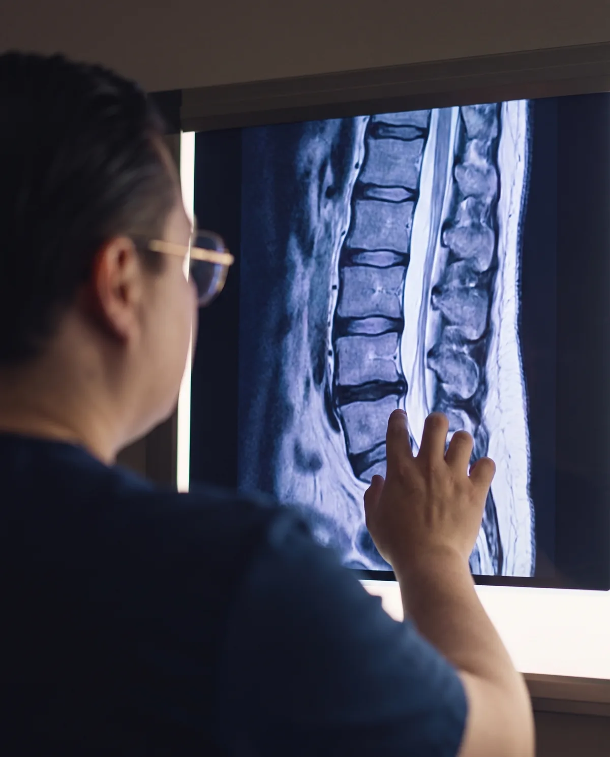
The physician team. Your spine MRI. A straight answer.

A patient coordinator collects your MRI. The physician team reads it and tells you plainly: candidate, not a candidate, or the surgeon is right. No pitch either way, and no second treatment sold before the first one has been followed up.

Book a call →
These therapies are not FDA-approved. Treatments are performed in Mexico by licensed physicians in a hospital setting. Individual results may vary. Not medical advice. Spine candidacy undergoes additional screening including neurologic assessment. Seek emergency care for sudden weakness, numbness, or loss of bladder or bowel control.