Medically reviewed by the Celva medical team · June 2026
The joint that
carries all of you.
For ankle osteoarthritis, plantar fasciitis, and chronic Achilles tendinopathy, MSC therapy is a precision-delivered, image-guided alternative to fusion, total ankle replacement, or repeated cortisone. Image guidance is non-negotiable in the ankle and forefoot.
Post-traumatic ankle arthritis is the dominant indication. Often a single old injury that drifted downhill.
When most foot patients first notice change. Slower than the knee because the joint loads every step.
You've been told
fusion or replacement.
For the ankle, the standard surgical answer is arthrodesis (fusion) or total ankle arthroplasty. Both are real options for end-stage disease. Patients with preserved tibiotalar joint space and a clear inflammatory driver are often worth evaluating first.
The foot and ankle are mechanical. Every step loads the joint, so the recovery curve is slower than knee or shoulder. The trade is durability, when therapy holds in this region, it tends to hold well. Ankle arthritis rarely sits alone: the tendons and ligaments that stabilize the joint are usually involved, and we treat those too, not just the joint surface.
If your foot & ankle surgeon has recommended fusion or replacement but your imaging still shows real joint space, an evaluation is a reasonable next step. We will read your films and tell you plainly.
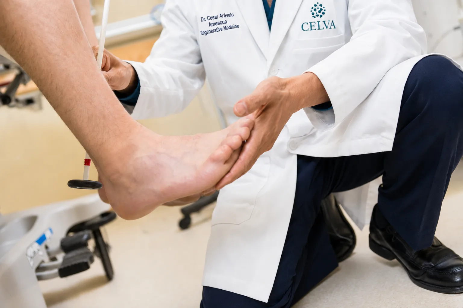
Ankle OA, preserved tibiotalar joint space
Post-traumatic or primary ankle arthritis with cartilage wear but limited bone-on-bone. Often a recommendation for fusion is already on the table.
Soft-tissue & peri-bony foot pain
Refractory plantar fasciitis, Achilles tendinopathy or tendinosis, and posterior tibial tendon dysfunction. Calcific deposits or heel spurs with chronic inflammation, after splinting, PT, eccentric loading, and cortisone or PRP.
End-stage ankle, severe deformity, neuropathy-driven pain
Bone-on-bone with significant varus or valgus malalignment, Charcot foot, or neuropathic pain, surgery or offloading is the correct tool.
Foot & ankle
we evaluate.
Every evaluation starts with a physician reading your imaging and history. Not every diagnosis qualifies. Candidacy is decided on staging, structure, and your specific case.
Ankle osteoarthritis
Post-traumatic or primary tibiotalar OA. Often a decade after an old sprain or fracture. Best response when joint space is preserved.
- Kellgren–Lawrence II–III
- Fluoroscopy or US guidance
- Subtalar joint, case-by-case
Plantar fascia, tendon & peri-bony
Ultrasound-guided injection for soft-tissue and peri-bony sites: refractory plantar fasciitis, Achilles tendinopathy and tendinosis, posterior tibial tendon dysfunction, and calcific deposits or heel spurs with chronic local inflammation. Where eccentric loading, ESWT, cortisone, or PRP have stopped holding.
- Plantar fasciitis, refractory
- Achilles tendinopathy / tendinosis
- Posterior tibial tendon dysfunction
- Calcific deposits / heel spurs
- Chronic foot inflammation
First MTP & midfoot OA
Hallux rigidus with preserved motion, midfoot osteoarthritis. Painful push-off, limited dorsiflexion, surgery framed as the only option.
- Hallux rigidus I–II
- Midfoot: tarsometatarsal joints
- US-guided intra-articular
How a foot session
actually runs.
The ankle and forefoot demand precision. Cells are delivered under fluoroscopy or high-resolution ultrasound, in-plane, with needle position confirmed before release. Off-loading instructions follow the patient home.
Joint cases like yours use bone-marrow and umbilical-cord MSCs, often in combination and sometimes with chondrocytes for cartilage support. The exact recipe, which cells, what doses, which routes, is decided by Celva's medical team after reviewing your imaging. Matched to your case, not pulled off a shelf. Why we use multiple cell types →
The entire procedure is physician-supervised at Hospital Angeles, Tijuana. Concierge transport from San Diego in the morning, discharge the same afternoon. A walking boot is provided for the first 48–72 hours when indicated.
Imaging & candidacy
Weight-bearing plain films and, where indicated, MRI read by the attending. Joint space, alignment, and structural integrity assessed. You'll hear either way.
Dose & targets
Cell type and dose, delivery route, and whether a same-day systemic IV is indicated.
Image-guided injection
Fluoroscopy for the tibiotalar joint, high-resolution US for tendon and forefoot. Needle position confirmed before release. Local anesthetic and twilight sedation (no general), so you stay comfortable and still and the physician can place the cells precisely.
Systemic IV
A second MSC dose IV in the infusion suite. Circulating cells are drawn toward inflammation and migrate toward the joints we just injected, reinforcing the local repair while calming inflammation across the foot and body-wide.
Scoring & check-ins
We score FAOS and VAS at baseline, then at 6 and 12 months, since the foot responds slower. A lighter pain-and-safety check comes around six weeks, and walking-protocol progression is confirmed at each visit. Repeat imaging on return for re-treatment.
What to expect.
And what we won't promise.
No outcome guarantees. What you get is a thorough evaluation, a realistic conversation, and treatment delivered to pharmaceutical-grade standards. Here is what our clinical follow-up generally shows for foot & ankle candidates.
Inflammation down
Morning pain shorter, swelling reduced. Loaded pain often unchanged yet, the joint carries every step.
Stride returns
Patients can walk further before pain. FAOS function improves for most qualifying candidates.
Peak window
The foot is slower than the knee. Most meaningful gains at 4–6 months. Some patients keep improving past that.
Durability window
Many foot patients hold meaningful results for one to three years, and often longer. Body weight, footwear, and activity load can shorten that window.
The foot and ankle take longer to respond than other regions. MSCs work through a slow paracrine process, and the joint never gets a real rest, you load it every step.
We measure progress on standardized scales: FAOS for function and VAS for pain, scored remotely at baseline and again at 6 and 12 months, with gait observation done in person on visit days.
End-stage tibiotalar arthritis with bone loss, severe varus or valgus malalignment, or neuropathic pain is generally not a strong MSC case. We'll tell you directly, and explain why fusion or replacement may be the right answer.
Foot-specific
questions.
Q.01 Can MSC therapy replace ankle fusion or replacement?
Q.02 How long before I notice improvement?
Q.03 Will plantar fasciitis really respond to this?
Q.04 Will I need a walking boot afterward?
Q.05 Bunions, hammertoes, or severe flatfoot?
Other joint regions
we treat.
Start with an
honest read.
A consult is a physician reviewing your imaging, history, and goals, then telling you plainly whether MSC therapy is a realistic option for your foot or ankle. If it isn't, we'll say so.