Celva / Treatments / Spine & joint pain, surgery avoidance

Medically reviewed by the Celva medical team · June 2026

§ 001 / Treatment · Spine & joint

Your spine, discs, and joints
deserve repair. Not replacement.

For patients facing joint-replacement surgery, spinal fusion, or told "there's nothing more to do," MSC therapy is a precision-delivered, image-guided alternative. Knee, hip, shoulder, hand, foot, plus neck, back & disc. It works with the body's own repair machinery, not around it.

Conditions
30+
Joint, spine, hand & foot indications we screen for
Delivery
2routes
Image-guided intra-articular and peri-articular + systemic IV, same session
Session
1day
Same-day return; an optional San Diego night is a personal travel choice, not required
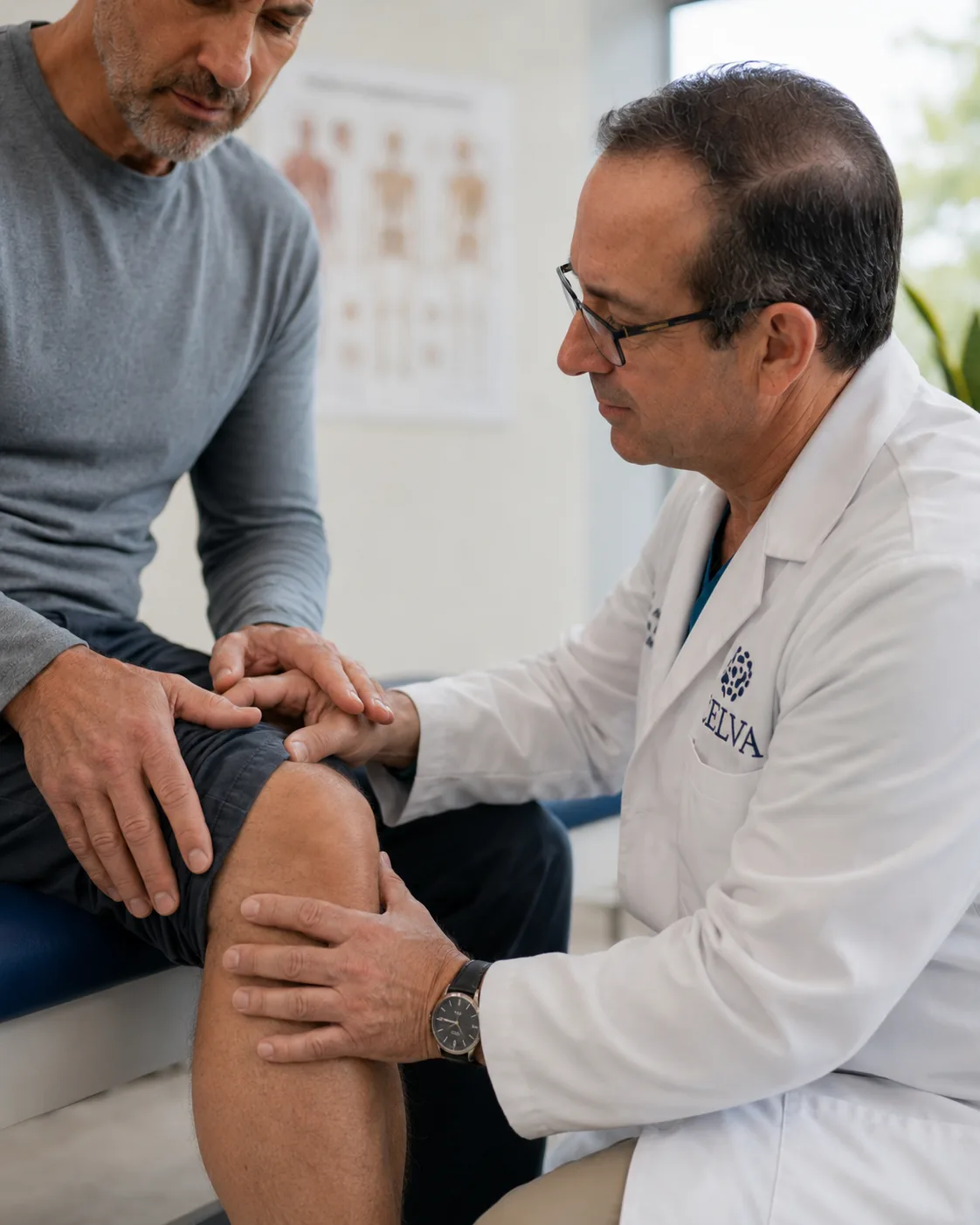
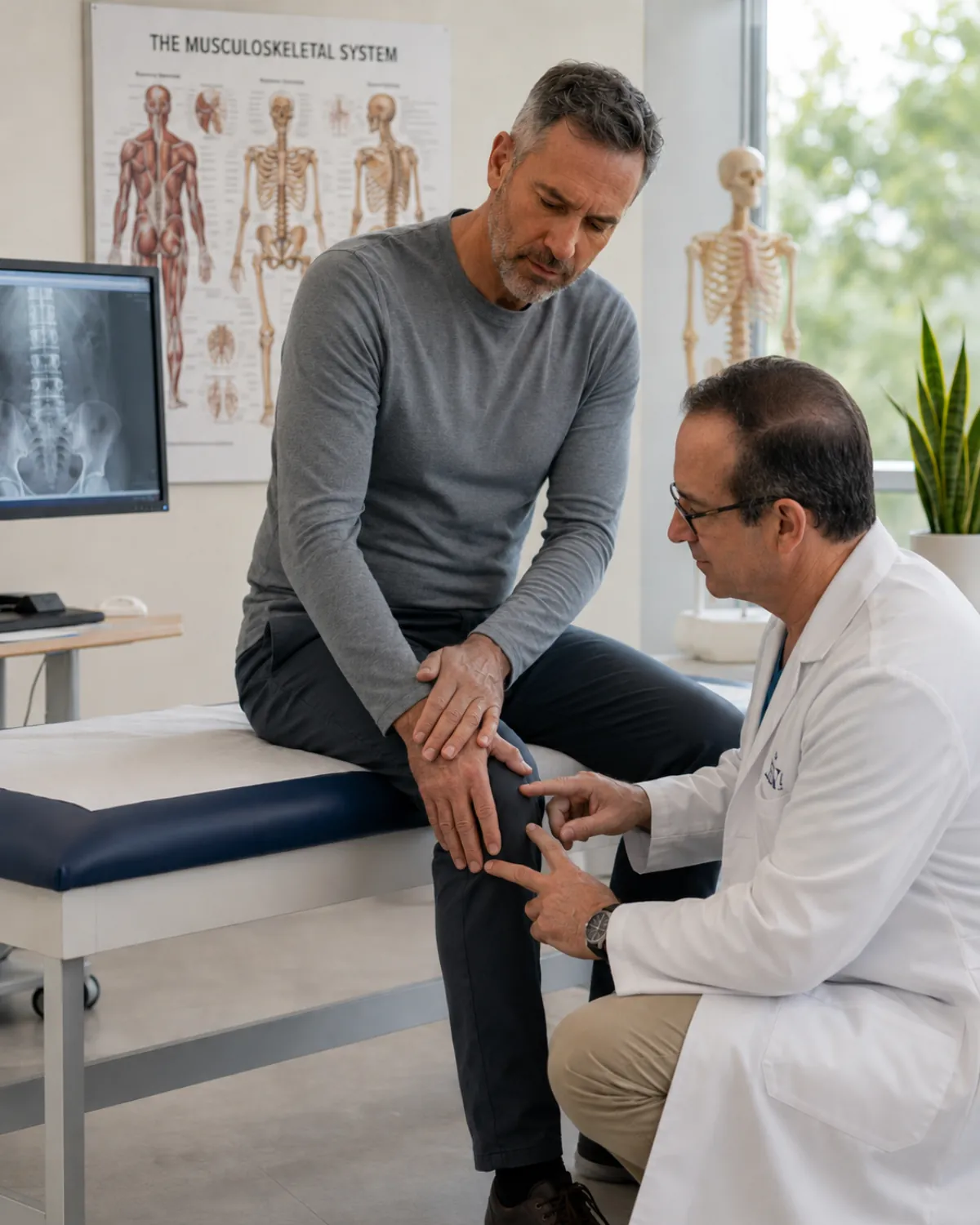
Physician evaluating a patient's knee in a clinical exam room
FIG. 01 · Region Atlas Knee · hip · shoulder · hand · foot · neck · back · disc
§ 002 / Indications

Six regions.
Thirty screened conditions.

Not every joint complaint is a candidate for cell therapy. We group by anatomical region and screen each indication against the evidence, your imaging, and your goals. Spine, hand, and foot work falls under the same image-guided injection standard as the larger joints. We treat each joint as a whole: the cartilage and the supporting ligaments, tendons, and muscles around it that drive much of the inflammation and pain, not the joint space alone.

Region · Spine
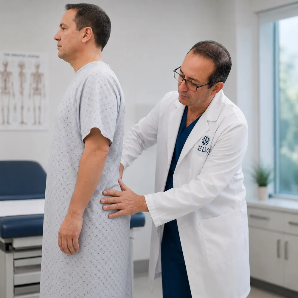
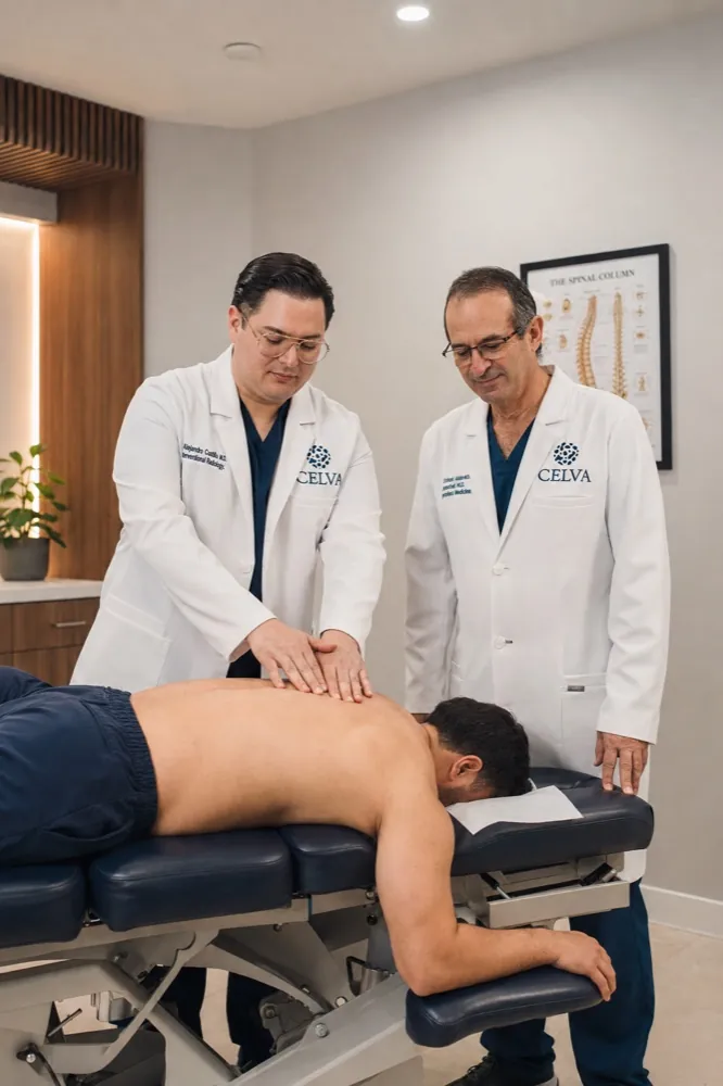
Two Celva clinicians evaluating a patient's spine on the exam table

The spine

Image-guided facet or epidural delivery. Disc therapy is case-by-case, with strict imaging screening.

  • Facet joint arthritis
  • Degenerative disc (screened)
  • SI joint dysfunction
  • Chronic neck or low back pain
  • Post-laminectomy pain
Learn more
Region · Knee
Physician examining a patient's knee

The knee

Our most-screened region. Image-guided intra-articular injection delivers MSCs directly to the joint space.

  • Osteoarthritis (I–III)
  • Meniscal tears
  • Patellofemoral pain
  • Post-surgical stiffness
  • ACL & MCL injuries
Learn more
Region · Shoulder
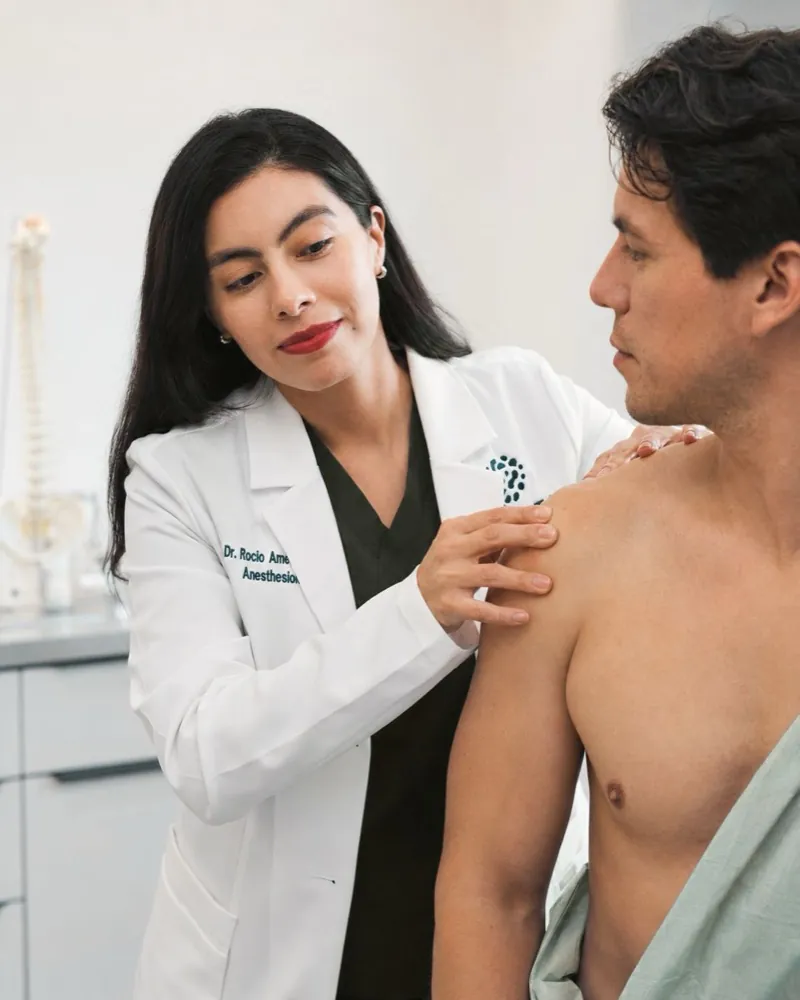
Celva clinician evaluating a patient's shoulder

The shoulder

Ultrasound-guided delivery to glenohumeral joint, rotator cuff tendon, or AC joint as indicated.

  • Rotator cuff tears (partial)
  • Adhesive capsulitis
  • AC joint arthritis
  • Biceps tendinopathy
  • Labral pathology
Learn more
Region · Hip
Physician examining a patient's hip during a clinical exam

The hip

Fluoroscopic guidance places cells precisely at the femoral head–acetabular interface.

  • Hip osteoarthritis
  • Labral tears
  • Avascular necrosis (early)
  • Hip impingement / FAI
  • Bursitis (recalcitrant)
Learn more
Region · Hand
Celva physician examining a patient's hand and thumb joint

The hand

Ultrasound-guided injection for thumb-base (CMC) arthritis and other small-joint OA. Same image-guided precision as larger joints, smaller anatomic targets.

  • CMC (thumb base) arthritis
  • Finger / IP joint osteoarthritis
  • Wrist osteoarthritis
  • Trigger finger (stenosing tenosynovitis)
  • Inflammatory hand pain
Learn more
Region · Foot
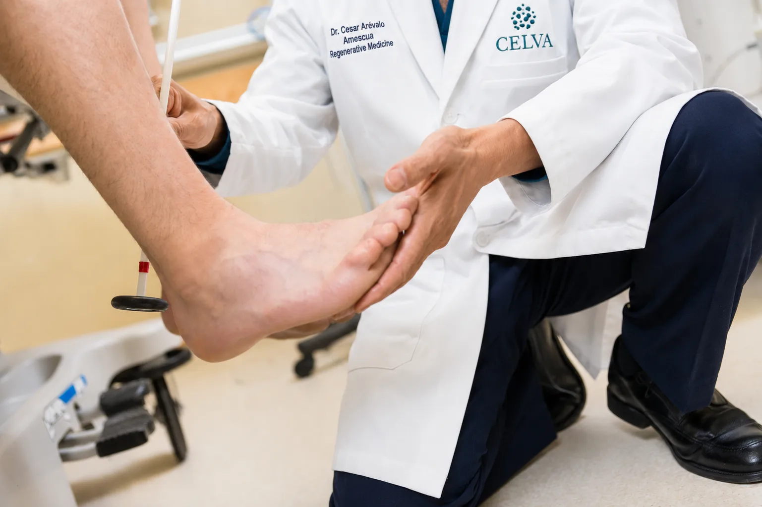
Celva physician examining a patient's foot for plantar and joint pain

The foot

Ultrasound-guided injection for plantar fasciitis, calcific deposits, and chronic foot inflammation. Targeted local delivery for soft-tissue and peri-bony sites.

  • Plantar fasciitis
  • Achilles tendinopathy / tendinosis
  • Posterior tibial tendon dysfunction
  • Calcific deposits / heel spurs
  • Chronic foot inflammation
Learn more
§ 003 / Candidacy

Who's a fit,
and who isn't.

A significant share of inquiries are declined by the physician team, not because we can't help, but because MSC therapy isn't the right tool for every problem. Here is the honest screen.

Likely candidate

You are a strong fit if…

  • You have recent imaging (MRI or weight-bearing X-ray), or you're open to imaging we perform on site at Hospital Angeles to establish a baseline.
  • Your pain maps to one or more joints or spinal levels we can target.
  • You've tried conservative care (PT, NSAIDs, corticosteroid or hyaluronic injection) and improvement plateaued.
  • You're facing a surgical recommendation (joint replacement, arthroscopy, discectomy) and want to delay or avoid it.
  • You have bone-on-bone (Kellgren–Lawrence IV) arthritis and want to delay or avoid joint replacement, with realistic expectations of a lower response than moderate cases.
  • Your BMI, medications, and systemic health allow an image-guided procedure.
Not a candidate

We would not recommend if…

  • There is active joint infection, uncontrolled systemic autoimmune disease, or recent malignancy.
  • You're in active oncology treatment (chemotherapy or radiation), or inside the standard surveillance window after a recent malignancy.
  • You're seeking a cure for a mechanical problem that requires hardware (unstable ligament, fracture, severe deformity).
  • You're pregnant, immunocompromised, or on anticoagulants we can't safely pause.
  • You're seeking a one-and-done fix without committing to the structured aftercare (rehab, follow-up calls, the rest).
§ 003.5 / Mechanism · local

What changes at
the injection site.

Chronically damaged tissue develops what we call a disrupted repair environment: persistent inflammation that prevents healing, fibrotic changes that replace functional tissue with scar, a local immune response that has become self-defeating, and compromised blood supply that starves the area of what it needs to recover. Image-guided injection places cells directly into that environment. The goal is to change the conditions so the body's own repair capacity can function again. That is the local half. A same-session systemic IV adds the other half: cells released into the bloodstream are drawn toward inflammation, so they migrate back toward the joint we just treated, reinforcing the local repair while calming inflammation throughout the body and supporting the broader benefits of a systemic infusion.

What changes at the injection site

Inflammation reduced at the source

Not managed systemically; reduced at the specific site where chronic inflammation is driving breakdown.

Fibrotic activity reduced locally

Scar tissue formation is reduced at the target site, helping preserve functional tissue where it is most at risk.

Local immune environment recalibrated

The immune response at the site, which in chronic conditions has become self-defeating, is brought back into a repair-supportive state.

Vascular supply supported

Cells support new blood vessel formation at the target tissue, contributing to perfusion that supports structural recovery.

Your protocol

The protocol, in cells.

No two joint cases get the same recipe. Bone-marrow MSCs by image-guided injection, umbilical-cord MSCs by injection or IV, and chondrocytes for cartilage support are all in the medical team's toolkit. Which cells, in what doses, by which routes is the clinical decision Celva's medical team makes after reviewing your imaging. Most joint cases use more than one cell type, delivered the same treatment day. Why we use multiple cell types →

§ 004 / Protocol

The joint protocol,
in specifics.

The general Celva methodology has five stages. For joint indications, here are the numbers that differ: dosing, route, session count, and what pre- and post-care look like.

Source
Multiple allogeneic cell types, matched to your case
Dose
Sized to the joint and your case
Delivery
Image-guided intra-articular and peri-articular (fluoroscopy + US)
Synergy
Systemic IV, same session
Sessions
One primary; booster at 6 mo if indicated
Anesthesia
Local; no general anesthesia required
Duration on site
~4 hours hospital-side
Return to activity
Off the joint 7–10 days, then graded build; load-bearing sport at 8–12 weeks
01 / Pre

Imaging review & labs

MRI and weight-bearing X-ray read by the physician team's attending. CBC, CMP, inflammatory markers, and coagulation panel within 14 days of procedure.

~2 wks pre-op · virtual
02 / Day-of

Image-guided delivery

Target joint is prepped under sterile technique. Cells are drawn into a 5cc syringe and injected under live fluoroscopic or ultrasound guidance. Position confirmed before release.

~45 min procedure · local anesthesia
03 / Synergy

Systemic IV infusion

A second allogeneic MSC dose, delivered IV in the infusion suite, monitored. This is where the two routes compound: circulating cells are drawn toward inflammation and migrate toward the joint we just injected, reinforcing the local repair while calming inflammation body-wide.

~1 hr · hospital infusion suite
04 / Post

Recovery & return

Monitored rest. Discharge instructions. Concierge transport back to San Diego once the attending physician has authorized discharge. You are walking the same day.

Physician-authorized discharge · same day
05 / Follow-up

Structured check-ins

Coordinator check-ins along the way, with formal scoring at baseline and again at 4, 8, and 12 months for most joints (6 and 12 for the slower-responding spine and foot). Pain-function scales (KOOS / HOOS / SPADI / FAOS / ODI) plus VAS, with repeat imaging on return for re-treatment.

Baseline + 4 / 8 / 12 mo · virtual
§ 005 / Clinical follow-up

Representative
joint cases.

De-identified, consent-verified cases from our clinical follow-up records. Chosen to represent typical trajectories, not only the best outcomes. Individual results vary.

Clinician placing an IV line for an MSC infusion
Knee · OA
Case PT-2401 · Knee OA III · 58 M

Replaced a recommended TKA with MSC therapy.

Patient was scheduled for a total knee arthroplasty after two years of conservative care and a corticosteroid course. Bilateral MRI showed Grade III chondral loss, medial compartment. Treated with image-guided intra-articular MSC plus systemic IV. Twelve-month re-image showed no further progression and KOOS function up from 42 to 76.

Baseline VAS
8 / 10
90-day VAS
3 / 10
180-day VAS
2 / 10
A patient swinging a tennis racket on a sunny outdoor court
Shoulder + Elbow
Case PT-2387 · Rotator cuff + lateral epicondylitis · 46 F

Tennis shoulder and elbow, back on the court.

Recreational tennis player with a supraspinatus partial tear and chronic lateral epicondylitis after PT, a corticosteroid course, and one PRP attempt. Ultrasound-guided MSC injection to the rotator-cuff tendons and the common extensor origin, plus systemic IV. Returned to baseline serve and groundstrokes at week 10 with no recurrence through one-year follow-up.

SPADI baseline
62
SPADI 90d
24
Return to sport
10 weeks
§ 006 / Next step

Every protocol is
built case by case.

There is no standard protocol for joint and spine cases. Celva's physician team at Hospital Angeles, Tijuana reviews your imaging, history, and goals, then designs the approach for your situation: which joint, single-region or bilateral, and whether an IV adjunct is indicated. The cells are matched to your case, not pulled off a shelf.

Start an intake and a Celva patient coordinator gathers your case for that review. Nothing is scheduled until the team has weighed in and you have decided to move forward.

Find out if you're a candidate →
§ 008 / Questions

Joint-specific
questions.

Q.01 If I'm scheduled for a knee replacement, is it too late?
Imaging tells us. Grade II and III are often strong candidates, even if surgery has been recommended. For end-stage Kellgren–Lawrence IV with bone-on-bone and subchondral loss, we'll still consider treating you if you want to delay or avoid replacement, just with a frank conversation about lower expected response. Send us your most recent MRI and we'll read it before your consult.
Q.02 Is it image-guided? Can it miss the target?
Every intra-articular delivery is performed under live fluoroscopy or ultrasound by an attending physician. Needle position is confirmed before cells are released. "Blind" injection (common at non-hospital clinics) is never used for joint work at Celva.
Q.03 Will it regrow cartilage?
We won't tell you that. MSCs are paracrine signalers. They release factors that modulate inflammation and support the body's own repair machinery. In joints, the typical observation is reduced pain, improved function, and slowed progression. Some patients show cartilage preservation on follow-up imaging. True regrowth is rare, and we don't lead with it.
Q.04 How is this different from PRP or a cortisone shot?
PRP concentrates your own platelets and growth factors. Cortisone is an anti-inflammatory steroid. MSC therapy introduces living, screened, expanded mesenchymal stem cells that signal for a longer therapeutic window. Three different tools. Many patients arrive having tried PRP and cortisone first, which is usually the right order.
Q.05 What does recovery look like for a joint case?
You walk the same day. Expect mild soreness at the injection site for 24–72 hours. No heavy impact or gym work for 7 days. Progressive return to full load at 6–8 weeks. Sport-specific return at 8–12 weeks depending on joint and baseline. You'll have a written protocol before you fly home.
§ 009 · Start here

See if you're
a candidate.

Send us your imaging, tell us about your joint. The Celva patient coordinator gathers your case and passes it to Celva's medical team, who tell you plainly whether MSC therapy is the right next step. If it isn't, they'll say so.

See if you're a candidate →
Not medical advice. Individual results vary. All patients undergo screening before treatment is recommended. Allogeneic cell therapies are performed by the physician team at Hospital Angeles, Tijuana, regulated by COFEPRIS in Mexico.