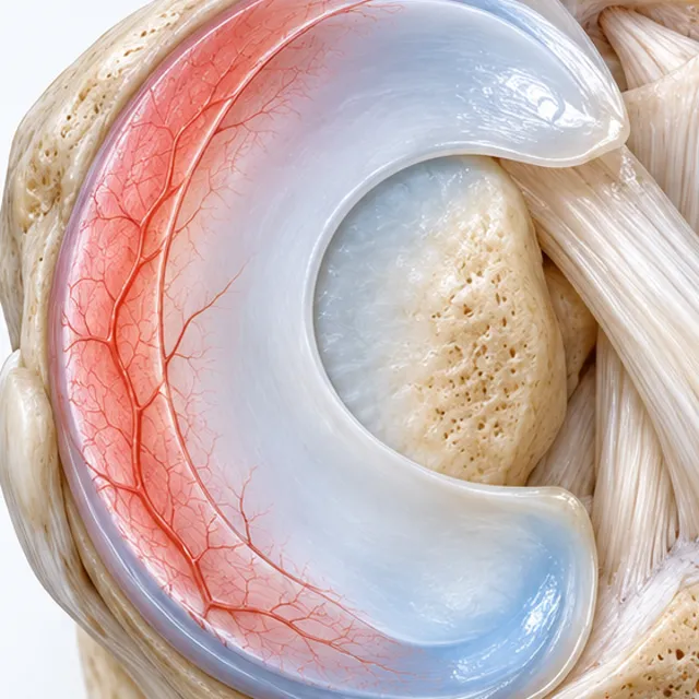
 Red zone
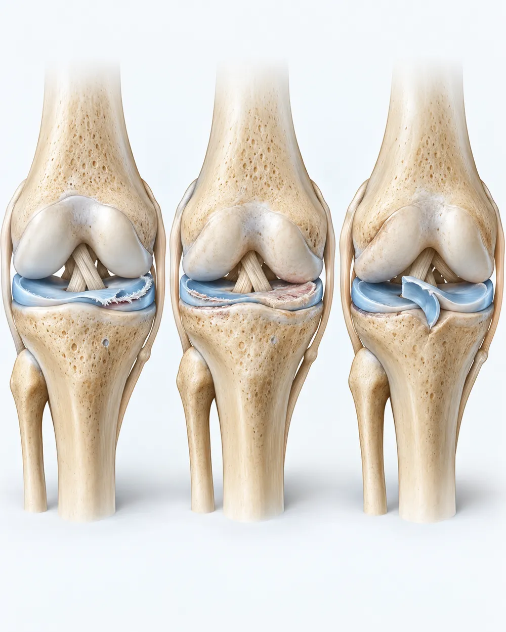
A red-zone tear at the outer edge can mend.
Blood reaches it, so a small tear can knit with rest and rehab, and a larger one can often be stitched (a meniscus repair).
You're probably reading this the night after the MRI, with "tear" in the report and an arthroscopy on offer. Whether it heals depends on where the tear sits and what shape it is. At Hospital Angeles in Tijuana, 25 to 30 minutes from San Diego, the physician team reads your MRI and says plainly which tear you have, and whether an image-guided injection fits it or a surgeon should take it.
Ten questions, no email, no call. The result names a sensible next step for your knee.
No meniscus is cut away. Cells go into the joint and the tissue around it, under live imaging.
Then physical therapy, and check-in calls at 1 week, then at 1, 3 and 6 months, then once a year, from home.
Each knee has two menisci, C-shaped pads of cartilage between the thigh bone and the shin bone. Only the outer edge has a blood supply: Arnoczky and Warren mapped it in 1982 (American Journal of Sports Medicine), vessels reaching the outer 10 to 30% of the medial meniscus and less of the lateral. That strip is the red zone; the rest is the white zone.
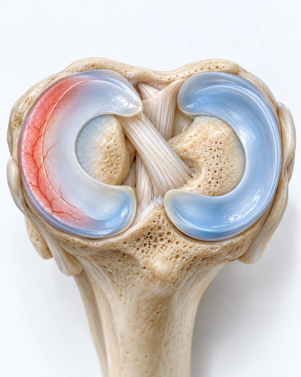 Red zone
Blood reaches it, so a small tear can knit with rest and rehab, and a larger one can often be stitched (a meniscus repair).
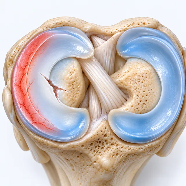 White zone
With no blood supply it has little to mend with. This is the tear most often trimmed.
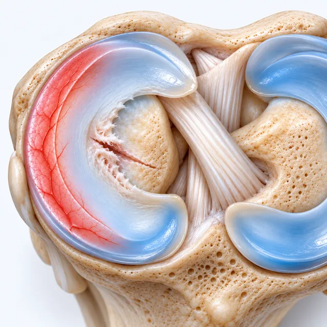 Degenerative
Wear rather than an injury, often with early arthritis. In Englund's 2008 study (New England Journal of Medicine), 61% of middle-aged and older adults with a tear on MRI had no knee pain that month.
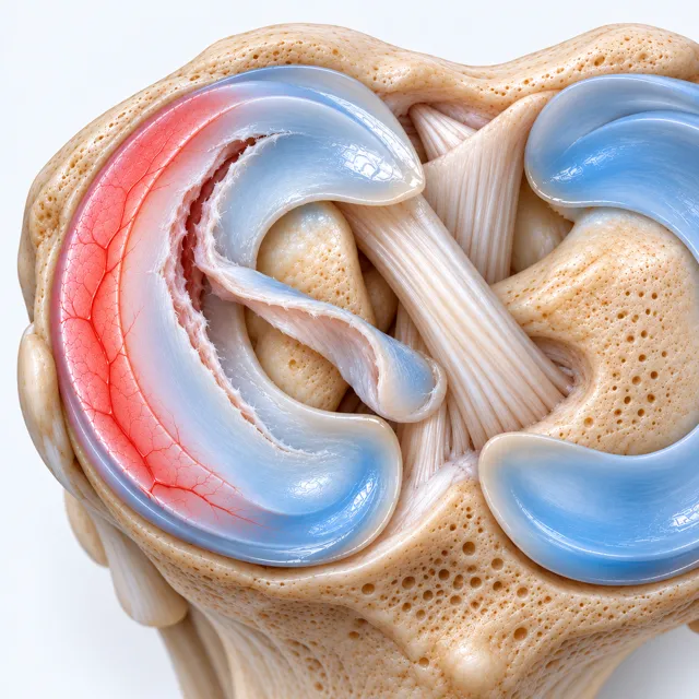 Bucket-handle
The knee catches or will not straighten: a mechanical block no injection moves.
Your MRI report names where the tear sits and its shape. Ten questions point you to a sensible next step for it.
Find your next step →"The other doctors told me that after physical therapy, if that didn't work for me, they only suggested surgery." That's Kathie M., 67, a Celva patient with a torn anterior meniscus. A tear that doesn't match where the knee hurts is a reason to keep looking, not a reason to inject.
Kellgren-Lawrence grade II to III (the standard X-ray scale for knee arthritis), joint space still there in at least one compartment, physical therapy behind you. The profile we accept most often.
Pain that physical therapy eased but never cleared, and an injection that wore off.
Wear that sped up after an injury or an arthroscopy. Cells do not regrow removed meniscus; the physician team reads what is left.
A knee that locks and will not straighten, or gives way from a torn ligament, is a surgeon's case, and we recommend one we trust. A knee that turns hot, red and swollen with a fever, or one you cannot stand on after an injury, is an emergency: go to an emergency room today.
Candidacy is confirmed from your imaging and the call. When you arrive, the physicians examine you in person to plan exactly how the treatment is done on your knee.
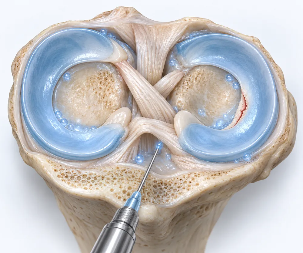
About 4 hours at the hospital, 15 to 30 minutes on the table, every placement under live imaging: fluoroscopy (a live X-ray) or ultrasound.
A Celva driver picks you up anywhere in Southern California and crosses the border with you. 1 or 2 companions are welcome, never required. The physicians examine the knee in person first to plan the injection.
Local anesthetic always, twilight sedation if you want it, and Dr. Rocio Ambrosio Nuño, anesthesiology, in the room.
Cells go into the joint space and into the tendons, ligaments and muscles around the knee, each placement confirmed on screen. They are designed to support that tissue, not to regrow a meniscus.
Bone-marrow and umbilical-cord MSCs (mesenchymal stem cells), sometimes with chondrocytes (cartilage cells), from a screened donor, grown in the on-site cGMP-compliant lab and released after 6 lab tests. Almost every joint patient also receives a systemic IV the same day.
Not sure your knee is this knee? Ten questions, no email, and the result names a next step.
Find your next step →Trimming (arthroscopic partial meniscectomy) is the operation most tears are offered. The trials below are why it is no longer automatic.
Watch Dr. Kirk Lewis's story"I ended up having surgery twice on my knee. I had torn meniscus in my knee."
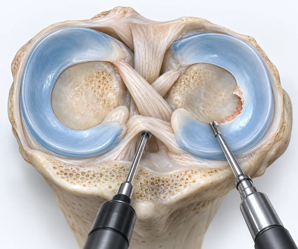
Katz, New England Journal of Medicine, 2013. 351 patients with a tear and mild to moderate arthritis: trimming plus therapy against therapy alone. Function improved about the same.
Sihvonen, New England Journal of Medicine, 2013. 146 patients with a degenerative tear: trimming against a sham operation. The trimmed knees did no better.
Kise, BMJ, 2016. 140 middle-aged patients: 12 weeks of supervised exercise against trimming. No clinically relevant difference.
Roos, Arthritis & Rheumatism, 1998. Knees whose meniscus was removed in an open operation, against matched knees: arthritis on X-ray about 14 times as common.
The honest limit: none of these trials tested cells. They show what trimming does for a worn knee, and why a 2017 guideline in the BMJ advises against arthroscopy for nearly all degenerative knees, not what an injection will do for yours. A locked knee, or a young knee with a tear that can be stitched, belongs to a surgeon.
No promises about outcomes. A schedule somebody keeps: four calls in the first 6 months, then one a year, from home, each measured against where you started.
Soreness for 24 to 72 hours, then 7 to 10 days of very limited activity before physical therapy. At 1 week your patient coordinator or aftercare specialist calls, by phone or video, and a physician joins when needed.
The cells work slowly, through signaling to nearby tissue (paracrine signaling). This call checks how you're settling. Treatment is one session, not a course.
The call asks about what you told us at the start: stairs, kneeling, getting up from a chair.
This call compares you with where you started. Whether a second treatment makes sense is decided at these reviews.
Every one of those calls is measured against the first one: the MRI and the knee you bring to it.
Find your next step →A coordinator typically calls within 1 business day, takes your history and tells you what to send.
Your MRI report and what has been tried.
Your MRI against where the knee hurts: candidate or not, and which tissue to treat.
A written answer, with a plan and an itemized quote for your case.
Steps 1 and 2 are the only ones you do. Take the call, send the report, and the rest is ours.
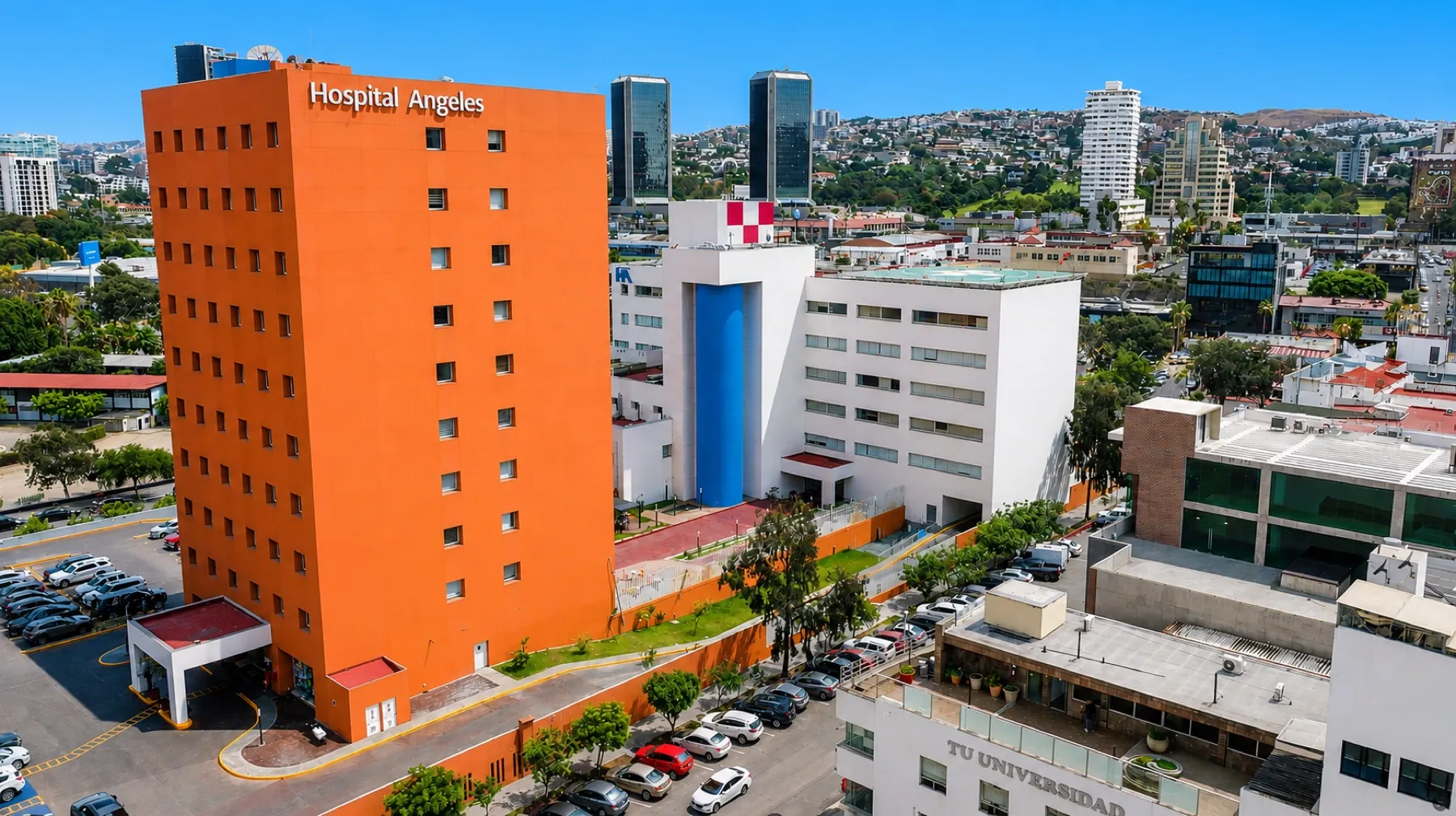
Book a call →Most cell therapy in Mexico happens in small, standalone clinics. Celva operates inside Hospital Angeles Tijuana, a 116-bed private hospital in the Angeles Health network, one of Mexico's largest. The physician team, the anesthesiologist and hospital imaging are in the building for your session.


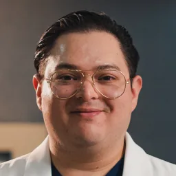

The physicians on every chart. Full bios →
Ask any clinic who is in the building during your injection. Then ask us.
Find your next step →Start with 10 questions, no email asked. When you want a person, the first call is with a Celva patient coordinator, about 15 minutes, and the physician team answers in writing: a candidate for cells, or a knee that belongs with a surgeon.